Supershift problem (EMSA problem) - I have a problem with supershifting my protein of interest (Mar/29/2006 )
Hi,
I have a problem with supershifting my protein in an EMSA reaction. I have tried all sorts of combinations (such as preincubation times, temperatures, reagent addition combination etc..). I don't see a slower migration of the band, or a diminishing of it.
I generally get the impression that lots of ppl have this problem(?), where they can see a gel shift however cannot do a successful supershift with antibodies they know that works (i.e. in western-blot, ICC, etc works). This is an extremely frustrating problem.
Therefore, I was wondering what the general consensus and opinion was in choosing a new antibody to buy. My current antibody is a monoclonal from chemicon. Should I try a poly clonal, or should I get a mono-clonal. Also, I have been able to to do my EMSA gel electrophoresis, and then transfer to a nitrocellulose membrane and conduct a western blot. This seems to work fine (especially with the antibody I'm trying to use for supershift).
I have also seen lots of papers where they haven't done a supershift. Is this worth the effort? Or is my approach to solve the problem incorrect?
Anyway, any opinions are welcome.
Thanks

A supershift is definitely worth the effort. Especially if it's critical to show a specific protein-DNA interaction. How else will people believe you otherwise?
Are you sure the epitope is adequately presented by the protein uner the EMSA conditions. It's possible that if you ran a denaturing western blot you'd get a band because the epitope is fully accesible. Don't forget that EMSAs are run in native conditions and so the epitope might be hidden in the tertiary structure of the protein. It's also quite possible that this specific epitope only become hidden upon binding to DNA (conformational change) which is why it works in other circumstances.
One way to check is to add a ridiculously large amount of antibody. How much are you using now? How much cell extract are you using? Is there published work of this supershift? If so then multiply the antibody concentration by 5 and then try. You are unlikely to get a nice band but you might get aggregation and the DNA complex remain in the well. This is still indicative of an antibody-protein interaction though.
As far as a new antibody....I would pick one with a different epitope (for reasons Doc Martin has admirably covered)
I also agree with him that adding a ton of antibody first is good; it takes more with supershift...relatively much more than doing a Western or anything else
one thing to mention...you have changed pre-incubation times. I have read that with some protein-Ab-DNA interactions, you want to add the extract to Ab first before adding your probe; others you want to add the extract to probe first before adding antibody...all depends on whether your antibody recognizes the DNA BP in the bound or unbound state (conformational changes occur upon binding; you see what I'm saying?) I do not know if you have changed the order of addition for your reaction components
Another thing...what supershift are you trying to show? If it's something like AP-1, for example, there are a whole host of proteins that can be involved in the binding, depending on the particular induction pathway...it will not always include cFOS, for example; sometimes the binding will be accomplished by MAF-ATF subfamilies instead, depending on upstream signaling events...even though it's AP-1, you won't ever see a supershift if you are probing with Fos...if you already know all this, I'm sorry to be loading it onto you????
good luck
Firstly, thankyou for your comments. Your feedback has been productive and well appreciated. Thanks again.
Generally I agree with u Doc, supershift is essential to demonstrate probe binding partners when done using a nuclear lysate. However, in my case I have bought a purified protein, which when analysed following SDS-PAGE and silver staining looks atleast 90% pure (it may be nearly 100%, but I’m being tentative).
I hadn’t considered the masking of the epitope as u suggested however. The antibody I used is a monoclonal, and according to the manufacturer it works for WB, ICC, IHC, in a variety of different species. The antibody is working under ICC and IHC conditions (which I have successfully tested; following paraformaldehyde fixing) suggests to me that antigenic epitope is not masked (however, I think paraformaldehyde does change conformation of the protein).
In regards to adding ridiculous amount of antibody, I have tried that too. I have mixed roughly 2.5ug of my protein of interest with increasing concentrations of antibody (1ug, 2.5ug, 5ug, and 10ug antibody). Still I do not get a banding profile representative of antibody binding protein (unless u think 10ug is not a ridiculous amount(?)-I don’t know).
There are no published examples of this protein being tested in a supershift assay- so there is no help there. Since this protein function is farely novel, there is only one paper where they have done an EMSA (no supershift, but published in JBC!).
I have tried a reverse order of reagent addition as u suggested (aimikins). This protein is predominantly present in the cytoplasm (greater than 85% cytoplasmic). Therefore, I doubt the antibody would be mainly binding a protein of DNA-bound conformation (I’m able to show cytoplasmic staining in ICC). If anything I would expect the antibody to be challenged by the DNA-bound form.
I am currently looking to purchase a polyclonal antibody (costs $500 Australian)! If this doesn’t pick up an epitope I don’t know. (I just had an idea, do ppl crosslink antibodies with a secondary in EMSAs?)
Another piece of information u might find useful is that the protein predominantly exists as a tetramer or dimer, and it is uncertain if it changes to a monomeric form upon binding. I’m not sure if this is a important consideration in gel shifts(?).
And, no aimikins I don’t want to get into AP-1 binding partners and dynamics (  ).
).
(There are a lot of AP-1 ppl in forums (Michael Karin would be very proud!))
anyway sorry for the long reply.
I look forward to any further suggestions.
Wow, I'm amazed you use that much purified protien to shift your probe. I'd have though a few tens of nanograms should have done it. You're using nuclear extract types quantities.
Anyway, what sort of percentage shift of the probe are you seeing with the 2.5 µg of protein? If it's a huge amount then you might find that you antibody is simply having difficulty shifting enough to make a difference. Also, you might try to expose your gels for a really long time.
I presume you using radiolabelled DNA? How are you visualising - X-ray film or Phosphorimager? A few years ago I wanted to see if I could elucidate protein binding at an initiator sequence in a TATA-less gene. Not an easy thing to do. After 48 hours of expose I saw nothing but after two weeks I could see a very weak band. Now whilst that result was still completely useless, it does go to show that exposing your gel for longer can be a good course of action.
I would suggest reducing the amount of purified protein to the lowest level you can that still gives you a decent shift. Then I'd add as much antibody as you think you can afford (10 µg seems like a lot to me - I've shown supershift/aggregation with 50-100 ng of antibody).
Is there any chance you could place an image of your unsuccessful gels on the forum? This might help.
I think Doc Martin is right. I usually use 2-10µg total nuclear extract but there's probably ng of the transcription factor of interest. Santa Cruz supershift abs are at 1-2µg/µl and they recommend using 0.5-1µl. As far as I know Santa Cruz and Active motif are a few of the only companies who test their abs in supershift.
Ceri
Firstly sorry for the very late reply.
I know this is a high amount of protein, but it is the quantity that gives me a reliable band everytime. Below I have shown a 'successful' gel (without supershift), and below that an antibody titration that has not worked:
Gel 1- lane1: probe alone; lane 2: GST-protein 2.5ug; lane3, 4, 5 purified protein of interest (with 0.5, 1.0 and 2.5ug protein respectively).The very dark bands on top are non-specific band where I think probe has got caugtht onto the well.![]()
Gel 2- lane 1-5 same as above; lane 6-9 2.5ug protein +increasing concentration of specifc monoclonal antibody (1ug, 2.5, 5, 10 respectively); lane 10-13 2.5ug protein + increaseing concentration of non-specific monoclonal anti-HA antibody (1ug, 2.5, 5, and 10 respectively).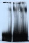
I do autoradiography and use films rather than phosphoimaging. Also, I thought antibody concentration would be more than enough at 10ug considering it is more than 1:1 molar ratio of protein to antibody.
Another question i have in regards to my EMSA is why the bands migrate lower and lower in the gel as I increase protein concentration?
Secondly I see in a lot of publications ppl displaying their 'free-probe'. Is this really important? (if not really I may run the gel for longer and get bands of interest lower)
My first comment would be that you have far, far to much probe in your assay. You can massively reduce the concentration of radiolabelled DNA. This will almost certainly help as you are increasing the ration of protein to 'receptor'. It will also make your images a lot clearer.
Second thing, in all of your lanes you still have a lot of material that hasn't migrated and has remained in the well. What is in your binding buffer?
Do you have carrier DNA? Do you have any detergent?
Use 1 µg poly(dIdC)-poly(dIdC) DNA per 20 µL reaction volume and add Triton X-100 to 0.1% final w/v. This will clean up your images.
Without ensuring 100% of your material has moved out of the well you cannot make any real conclusions. How do you know what species are represented in that band? It may be supershifted DNA, impossible to tell as it is.
As for including the free probe - I am totally in favour of showing the entire gel (from wells to probe). Were I reviewing a paper in which only the shifted band was shown (as in many Western blot images) I would demand that the whole gel was shown as it is not possible to rule out manipulation of the image and/or selective editing. It mean you could leave out all the non-specific rubbish going on.
Also it looks as though you are getting quite a lot of uncomplexed ssDNA. Check you annealing protocol. It might not be working efficiently.
Here is an example of how clean you can make an EMSA. This is a supershift expt from my thesis. I have broken the rule I now follow and have cropped the bottom of the gel. However, there was no ssDNA and the image shows nearly the entire band (maybe only 10-15% chopped off). Notice how much more defined the free probe band is. You can massively reduce your free probe conc.
This also make the shifted bands far more deifined as you get less background and smearing. You can also use less extract which minimises lanes overloading and smiling.
I would like to add one thing....sometimes the probe-antibody-protein complex is inhibited from entering the lane during electrophoresis, and you can sometimes improve the problem by adding a small amount of BSA to your binding buffer as well
Doc Martin has raised many excellent points