How to view the encapsulated cells under microscope? - (Feb/05/2014 )
Dear all,
Did anyone have any idea how can I view the encapsulated cells under light microscope? How can I make the protective layer become more visible so that I can compare the differences between non-encapsulated and encapsulated cells? Thank you
What are encapsulated cells?
The bacterial cells that being encapsulated by a protective layer, such as maltodextrin and gelatin.
Use India ink prep to see the capsule.
https://web2.uwindsor.ca/courses/biology/fackrell/Methods/Capsule_Stain.htm
You can typically remove the capsule with a dilute solution of NaEDTA.
The classic stain for capsules is rose-bengal followed by nigrosin, the bugs will stain bright pink and the capsule will appear as a clear ring around the bacteria against the dark background.
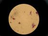
Thanks for the protocol. I was using Anthony's staining method to view the encapsulated bacterial cells. I have a question that would like to seek for your advice. Attached is the cells image after staining with crystal violet.
First image (IMG_6985) is the bacterial cells incubated with maltodextrin and fructo-oligosaccharide. Did anyone know what is that pinkish stuff? Is maltodextrin or fructo-oligosaccharide? Or the combination of both of them?
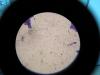
Second image (IMG_7007) is the bacterial cells incubated with maltodextrin only. I noticed there is a number of sphere shape capsule-like compound. Is that the encapsulated bacterial cells?
Thank you.