Need help on STAT1-GFP nuclear translocation - (Jan/25/2013 )
Hello! Everyone,
(1) I am using a cell line expressing STAT1-eGFP to demonstrate the nuclear translocation of STAT1-eGFP after IFN-treatment.
My purpose is to compare the efficacy of IFNs with this system.
Briefly, HEK293 (or HELA) cells were transfected with a plasmid STAT1-eGFP. (The plasmid should be fine because it was bought from Addgene, and other published studies have used the same plasmid for the same nuclear translocation experiment.)
I have transitorily transfected cells, and I also have HEK293 stably expressing STAT1-eGFP.
STAT1-eGFP located in cytoplasm that is consistent with others’ researches.
Then I treated those cells with 1000U/ml IFNbeta or IFNalpha. However, I have not observed the accumulation of STAT1-eGFP in nuclear after 30min, 1hours, 2hours, 4hours, 8hours, and 12hours post-treatment.
IFNs should be fine. I have used them for IFN-receptor internalisation experiment. They worked very well.
I do not understand why there was no nuclear trafficking.
(2) I have also used immunofluorescence to detect the phosphorylation state of STAT1.
Firstly, HELA (HEK293, Raji and U973) was cultured on slide, then cultured in medium with 1% serum 24 hours before IFN-treatment. 1 hour before IFN treatment, cells were cultured in medium without serum.
After IFN-treatment, cells was washed, fixed and permeabilized (I have tried Paraformaldehyde 4% for fixation then 1% saponin; or cold methanol treated at -20C for 10min). After washing, rabbit anti-STAT1-p (T701) (from Cell signalling) was used as 1st antibody, then goat anti rabbit antibody-Alexa594 (I have used whole goat IgG and also its F(ab)’2; from Cell signalling) was used as 2nd antibody.
However, I did not detect any specific signalling of STAT1-p.
The last possibility that I will check is the serum for cell culture medium. My colleagues also have a bit problem with this serum when doing cytokine related experiment. However, I have starved cells before IFN-treatment. I am very confused.
Any suggestion and help is appreciated!
Thanks a lot!
Based on what you've said, there is no obvious answer. Can you show some images?
Thanks a lot!
The image is HEK293 cell (transitorily transfected with plasmid expressing STAT1-eGFP https://www.addgene.org/12301/) after IFN-treatment (1000U/ml). The location of STAT1-gfp is the same as before the treatment.
What I expect is something like this one
https://www.ncbi.nlm....2181/figure/f3/ ((A), 2nd image=Vector+ IFNbeta)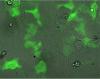
In this image, Hela cell cultured on cover slide (starved by using DMEM + 1% serum 24 hours before treatment, and no serum DMEM 1 hour before treatment) was treated with 250mp IFNbeta for 25min, then washed, fixed with cold methanol for 7min at -20C; after washing, anti phospho-STAT1antibody (https://www.cellsignal.com/products/9167.html) was added. 2nd antibody was goat anti rabbit F(ab)'2-Alexa594 (also from Cellsignaling).
When taking this confocal picture, the voltage was quite high otherwise we can see nothing. Hence, the red color here is just autofluorescence.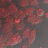
Is the phospho stat-1 stuff being done on the same GFP plasmid as the localization stuff?
If so - check the sequence of the plasmid to ensure that it has the correct Stat-1 sequence (mistakes can easily be made - you should do this for all plasmids entering the lab).
Could it be that your IFN is degraded in some manner, which would result in you not seeing the effect you expect?
I agree that non-functional IFN could explain both results. Try with a fresh batch and do a western blot in parallel to be certain that the IFN is working. The phospho-Stat1 antibody you are using is absolutely amazing. I've used it for IF and flow cytometry ant the signal to noise is wonderful.
The phospho-stat1 translocation experiment was done with non-transfected Hela cell.
IFN should be OK. I have tried fresh stock of IFN and both IFN alpha and beta. I have worked with the aliquot of same IFN in other experiment that worked well. Hence the quality of IFN should be fine.
Hello! Doxorubicin,
I am not very experienced in intracellular staining.
To fix/permeabilize protein in nuclear, which method is better? Cold methanol at -20C for 10 min (I have red this method on several publications), or PFA 4%+ saponin 0.5% (this is rather for cytoplasmic protein)?
Which cell did you use for your IF experiment of phospho-stat1? I have use Hela and Hek293. I think that the 2 cell lines should have typical Jak/stat(1) signaling pathway.
Is WB more sensitive than IF? I find IF is pretty tricky.
IF should be pretty straightforward and sensitive. I always do 10 minutes of 3.7% PFA at 37C followed by at least 30 minutes of -20C methanol (100%). This should be fairly similar to the standard Cell Signaling Technology protocol they recommend on their website. Find their protocol and follow it carefully.
WB is not necessarily more sensitive than IF...it's just the only raw data that anyone presents these days, so it is easy to look at and tell just what is working/not working....but if your lab doesn't do western blots, then it's likely too much trouble.
HeLa and 293 cells should respond well to IFN. 293 cells, depending on the line, will often jump off the slide during PBS washes, so are sometimes not ideal for imaging. I use WISH cells, which are similar to HeLa (or identical if you believe ATCC).
Should I starve the cells before IFN treatment in WB experiment? In publication it was not mentioned, hence I guess that it is not necessary. On the contrary, to stave the cells before IFN treatment seems necessary in IF experiment. Am I right?