dot blot - (Sep/29/2011 )
I am trying to explore the expression of an HA-tagged protein in cell lysate.
I used dot blot but there was a problem. Dots did not penetrate the PVDF membrane easily and formed a glassy layer after about 4 hours. On the other hand some dots became transparent like an oil spot.
Blots became white (not dark) in the developed film. I also had a dot from my primary antibody as a control which became dark in contrast to samples.
Does anybody have a suggestion?
Did you wet your membrane in 100% methanol before blotting? PVDF goes translucent/transparent when wet, so the dots going like an "oil spot" may very well be the only ones that have actually been absorbed by the membrane.
When you say the blots became white (not dark), do you mean the whole film, or the dotted parts? If the first, that probably means your dot blot has not worked at all, and there's nothing there to show black. If the second, your dots are white into a darker background, that could be explained by too much protein.
Could you tell us a bit more about what exactly is that you did and maybe post an image? It'll be easier for us to help you.
Thanks for your reply.
Should I activate my membrane before performing dot blot?
I drew 3 lines with pencil on the membrane for separating 6 regions. I put about 5 micro liters of each sample on the dry PVDF membrane and waited for dots to dry.
my oily dots were the middle-up and down-right in the attached picture. They lack my protein of interest!
About primary antibody dot (up-right) I made a mistake. my dot is the white spot. I am not sure what the dark spots are.
The other three dots were not absorbed easily, I scattered them around using pipette tip and saw that they became sticky. These dots were reflecting light when I started blocking the membrane.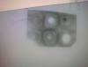
Yes, you need to activate the membrane. PVDF is highly hydrophobic and will not bind your proteins otherwise. Soak it in 100% Methanol and then in water or wash buffer. I'd also recommend to blot your protein while the membrane is still wet, then allow to dry.
So you could draw your lines in pencil, then soak in methanol, followed by water or buffer. After this let the excess liquid off but DO NOT LET THE MEMBRANE DRY. Place the membrane on top of a bit of cling film, saran wrap or parafilm and do your blotting. Then allow to air dry for the proteins to be fully absorbed in the membrane.
Finally, I think 5ul is a bit too much. If you want to blot that much, I would suggest adding 1ul at a time.
Alternatively, you could use nitrocellulose which will absorb your proteins much better and will solve the hassle of pre-wetting and keeping wet.
Not sure how to interpret your results. I think the "oily spots" are a result of some protein dried off in the surface of the membrane (not properly absorbed), and all the blackness is high background due to poor blocking and washing but I don know how to explain the white spots. 
what is your full protocol?
Here are a few links that will help you:
https://www.bio-rad.c...in_4006127A.pdf
https://www.millipore...ech1/tn1004enus
This one is for Nitrocellulose, but you can do the same with PVDF by previously soaking in methanol and water.
https://www.abcam.com...%20protocol.pdf
Good luck!
Thank you so much.
I read different protocols including:
https://www.rndsystems.com/rnd_page_objectname_wb_dotblot.aspx
https://westernblot.org/dot-blot/
https://biologicalworld.com/dotblot.htm
https://www.abcam.com/index.html?pageconfig=resource&rid=11452
https://www.westernblotting.org/dot%20blot%20protocol.htm
https://www.genetex.com/Images/Protocol/Dot%20Blot.pdf
Thanks again
Hope being in contact after my next dot blot