Western blot: problem with protein migration - trouble shooting......... please help! (Jul/05/2010 )
Hi everyone,
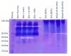
I’ve been doing Western blots for about a year with no problems. I recently went to a new lab and despite using the same recipes for EVERYTHING, I cannot get my Western blots to work. The primary problem appears to be the migration of my total protein extract (from HeLa cells) through the SDS-PAGE. The marker runs beautifully, but after staining the blot with Ponceau, the total protein appears impeded and doesn’t migrate much farther than 80 kDa. It’s not a problem with transfer because Coomassie stained gels have the same problem. I’ve also tried a number of different Laemmli buffers to no avail so that doesn’t seem to be the problem either.
However, when I run purified BSA and stain with Coomassie, I get a band at the appropriate size (~66 kDa). I don’t think it’s the gels. I thought it might be the protein extraction so I tried using M-PER (from Pierce) and RIPA (from Millipore) and get the same results.
I have no idea what to try next. Any suggestions would be fantastic. I have my solutions listed below and a PDF of my protocol attached. i also attached a picture of a Coomassie stained gel. All samples loaded are 20 ug of protein.... I probably should have ran 10 ug, but oh well.
Thanks.

My solutions.....
----------------------------------------------------------------
Buffer Solutions:
1X SDS Running Buffer:
100 mL 10X running buffer
900 mL dIH20
1X Transfer buffer:
100 mL 10X transfer buffer
100 mL methanol
800 mL dIH2O
1X TBST
100 mL 10X TBS
10 mL 10% Tween-20
890 mL H2O
---------------------------------------------------------------
Other Solutions:
Upper gel
4X Stacking gel buffer:
0.5M Tris, pH 6.8 (15.14g into 240 mL dH2O)
0.4% SDS (from 10% SDS=10 mL)
Autoclave PRIOR to adding SDS.
Lower gel
4X Separating gel buffer:
1.5 M Tris, pH 8.8 (45.42g into 240 mL dH2O)
0.4% SDS (from 10% SDS=10 mL)
Autoclave PRIOR to adding SDS.
10X SDS Running Buffer
1.92 M Glycine (288 g)
250 mM Tris (60 g)
0.1% SDS (10 g)
Make up glycine and tris in 2L of dIH2O and pH to 8.3. Autoclave. When the solution is cool, stir in 10 g of SDS.
10X Transfer buffer:
1.92 M Glycine (288 g)
250 mM Tris Base (60.6 g)
Make up in 2L of dIH2O and autoclave.
10X TBS
1.5 M NaCl (175.32 g)
0.5 M Tris (121.14 g)
Make up in 2L of dIH2Oand pH to 8.0. Autoclave.
10% APS
Make up a 1 g APS in 10 mL sterile dIH20 and divide into 0.5 mL aliquots.
Store at -20˚C.
As APS degrades, it begins to acidify. To test the integrity of the APS aliquot, spot onto pH indicator paper. If the solution soaking into the paper does not change the color to acid past the initial spot, then the APS has not degraded.
Ponceau S Dye
(0.1% Ponceau in 10% acetic acid)
Dissolve 0.5 gram of Ponceau in 400 mL dIH2O
Add 50 mL of glacial acetic acid
Qs to 500 mL
Just a guess....but it looks like you forgot to add the SDS (or maybe reducing agent) to your Protein Sample Buffer.
I wish it were that easy. The B-mercaptoethanol has been added. I've even tried the Biorad Laemmli. I thought it might be the sample buffer as well, but I think I've ruled it out.
I also remade all the solutions to ensure SDS was added.
Hi..
Could you please help me with western blotting for Collagen II?
Should I check for the protein in the medium or cell layer?
I am culturing on 24well plates and I want to check for collagen II presence.
Please help me....
Rachel USF on Jul 5 2010, 01:39 PM said:
I’ve been doing Western blots for about a year with no problems. I recently went to a new lab and despite using the same recipes for EVERYTHING, I cannot get my Western blots to work. The primary problem appears to be the migration of my total protein extract (from HeLa cells) through the SDS-PAGE. The marker runs beautifully, but after staining the blot with Ponceau, the total protein appears impeded and doesn’t migrate much farther than 80 kDa. It’s not a problem with transfer because Coomassie stained gels have the same problem. I’ve also tried a number of different Laemmli buffers to no avail so that doesn’t seem to be the problem either.
However, when I run purified BSA and stain with Coomassie, I get a band at the appropriate size (~66 kDa). I don’t think it’s the gels. I thought it might be the protein extraction so I tried using M-PER (from Pierce) and RIPA (from Millipore) and get the same results.
I have no idea what to try next. Any suggestions would be fantastic. I have my solutions listed below and a PDF of my protocol attached. i also attached a picture of a Coomassie stained gel. All samples loaded are 20 ug of protein.... I probably should have ran 10 ug, but oh well.
Thanks.
My solutions.....
----------------------------------------------------------------
Buffer Solutions:
1X SDS Running Buffer:
100 mL 10X running buffer
900 mL dIH20
1X Transfer buffer:
100 mL 10X transfer buffer
100 mL methanol
800 mL dIH2O
1X TBST
100 mL 10X TBS
10 mL 10% Tween-20
890 mL H2O
---------------------------------------------------------------
Other Solutions:
Upper gel
4X Stacking gel buffer:
0.5M Tris, pH 6.8 (15.14g into 240 mL dH2O)
0.4% SDS (from 10% SDS=10 mL)
Autoclave PRIOR to adding SDS.
Lower gel
4X Separating gel buffer:
1.5 M Tris, pH 8.8 (45.42g into 240 mL dH2O)
0.4% SDS (from 10% SDS=10 mL)
Autoclave PRIOR to adding SDS.
10X SDS Running Buffer
1.92 M Glycine (288 g)
250 mM Tris (60 g)
0.1% SDS (10 g)
Make up glycine and tris in 2L of dIH2O and pH to 8.3. Autoclave. When the solution is cool, stir in 10 g of SDS.
10X Transfer buffer:
1.92 M Glycine (288 g)
250 mM Tris Base (60.6 g)
Make up in 2L of dIH2O and autoclave.
10X TBS
1.5 M NaCl (175.32 g)
0.5 M Tris (121.14 g)
Make up in 2L of dIH2Oand pH to 8.0. Autoclave.
10% APS
Make up a 1 g APS in 10 mL sterile dIH20 and divide into 0.5 mL aliquots.
Store at -20˚C.
As APS degrades, it begins to acidify. To test the integrity of the APS aliquot, spot onto pH indicator paper. If the solution soaking into the paper does not change the color to acid past the initial spot, then the APS has not degraded.
Ponceau S Dye
(0.1% Ponceau in 10% acetic acid)
Dissolve 0.5 gram of Ponceau in 400 mL dIH2O
Add 50 mL of glacial acetic acid
Qs to 500 mL
Hi,
I didnt check well your protocol! but, just by reading thay you used ripa buffer, and Hela cells. i would like to participate to your discussion modestly.
Im using ripa buffer as well to solubilize my protein of interest, which is expressed in HEK cells.
I also, have the problem before, to see huged band of proteins sticked at the top of the gel, after several trials, it was problem of the proteins that aggregate.
So, why don't you solubilize better your extracts before loading SDS-PAGE gel. in other words, be careful to work on ice, if u use ripa buffer. and after solubilization, leave the samples at 4°C on rotatin wheel, for 20 to 30 min.
then, quantify your proteins well. and also, may be you have to boil for 3 to 5min, before loading the samples!
hope this can help.
good luck
Could it be that your total protein extract are aggregating together and forming dimers? Maybe you should heat your sample lysate and sample buffer for 10 minutes at 70*C rather than 5 min at 95*C
and for 'scarlet'- its rude to hijack someone's thread- make your own.
Chiapet974, I'll try the heating at 70*C for 10 min.
And luciana, the way I extracted the proteins was I collected the cells by scraping with PBS, removed the supernatant, added RIPA or M-PER + protease inhibitors, let sit on ice for 15 min with occasional tapping, then centrifuged and collected the supernatant. You think rotating will help? I do agree that it must be the extraction, though.
Chiapet874 on Jul 6 2010, 05:30 AM said:
and for 'scarlet'- its rude to hijack someone's thread- make your own.
do you remove the extraction buffer (by precipitation, dialysis, or whatever) before adding laemmli sample buffer?
if not then you probably have too much salt in the sample
and
nonionic surfactants, such as triton and nonidet, compete with sds. if the concentration is high enough then the sds may not bind to or will be stripped from the protein.
mdfenko on Jul 7 2010, 11:08 AM said:
if not then you probably have too much salt in the sample
and
nonionic surfactants, such as triton and nonidet, compete with sds. if the concentration is high enough then the sds may not bind to or will be stripped from the protein.
mdfenko: Do you recommend that I do an acetone precipitation on the protein and then reprecipitate in an SDS based buffer?