Detecting phosphorylated Erk1/2 - (Mar/12/2010 )
Hey people,
I am trying to detect phospho-Erk1/2 in cultured MDCK.1 cells. I used EGF stimulation as a positive control and I did 24h serum starvation before stimulation. Now the problem is that I can only detect p44 phosphorylation and not matter how hard I bit MDCK cells with EGF I still got phospho-p44. It's quite a bizzare as the antibody I used works perfectly in other cell lines.
Does anybody have any clues for what might cause the problem?
Eric
Eric on Mar 12 2010, 12:56 AM said:
I am trying to detect phospho-Erk1/2 in cultured MDCK.1 cells. I used EGF stimulation as a positive control and I did 24h serum starvation before stimulation. Now the problem is that I can only detect p44 phosphorylation and not matter how hard I bit MDCK cells with EGF I still got phospho-p44. It's quite a bizzare as the antibody I used works perfectly in other cell lines.
Does anybody have any clues for what might cause the problem?
Eric
No one has any idea?
Are you using phosphatase inhibitors?
Halfro22 on Mar 25 2010, 01:11 PM said:
Yes, otherwise I may not be able to detect phospho-p44. The weird thing is just I can not detect phospho-p44 in MDCK cell lysate while both p42 and p44 were detected when using VSMC lysate.
I've just started working with p42/p44 and phosphorylation in Jurkat cells and my western blots seem to show only phosphorylated p42 (I think it's p42 and not p44). If I overexpose the blots and background starts to show up, I can faintly see a band where p44 should be but I also see faint bands of multiple different sizes showing up. Any idea what is going on? Are these other bands related to the p44/p42 sequences??
yes this is due to sequence similarities. anti-phospho-antibodies are often raised against short synthetic phosphorylated peptids. sequence should be mentioned in data sheet. so what you see are erk-precursors and proteins sharing sequence motifs and on.
to be sure about p44/p42 phosphorylated or not, treat blot-membrane with a anti-ERK1/2 antibody. you should see two bands, of course. If not, p44a and p42 are probably together in same band. use more harsh denaturating agents or change composition of your separating gel for better protein separation.
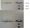
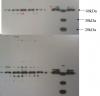
Thanks! I've attached a picture of the blot, one exposed for 2 minutes, the bottom for 4 minutes, and I'm probing against anti-phospho p42/p44 (aka MAPK Erk1/2) (4-12% Bis-tris gel).
Do you think that they are resolving as the bright single band in most of the lanes? (The difference between the lanes is just different cell treatments) Or do you think it could be either the higher band or lower band I indicated with the red star? It doesn't seem to make sense that it would only show up in some lanes and not others if the other band is there, correct?
Anyone have any recommendations for how to resolve the two bands using the Novex/invitrogen system?
Since I'm new to MAPK signalling work, I was wondering if anyone has any good recipes or recommendations for lysis buffers. I also want to look at membrane proteins in the same sample- and do people typically include the nucleus in their cell lysate, as some signalling molecules translocate to the nucleus, correct?
Thanks again!